

Key takeaways
- A CBCT scan is a 3D dental X-ray: the scanner makes one quick rotation around your head and a computer builds a three-dimensional picture of the teeth, jaw and surrounding structures.
- It shows what a flat 2D X-ray cannot: bone height, width and density, the exact position of nerves and sinuses, the shape of root canals, and unerupted or impacted teeth.
- It is genuinely useful for specific jobs such as planning implants, assessing bone before a graft and difficult wisdom teeth; routine check-ups and simple fillings do not need one.
- The dose is higher than a single small dental X-ray but still low, so a careful clinic only takes a CBCT when it will genuinely change the plan.
Most dental X-rays are flat, two-dimensional pictures, and for everyday dentistry that is exactly what is needed. Sometimes, though, a dentist needs to see in three dimensions: how much bone there is, where a nerve runs, or how a hidden tooth is lying. That is what a CBCT scan provides. If you want the background on the routine digital X-rays used at most visits, our guide to what an RVG dental X-ray is covers that ground; here we look at the 3D scan and, just as importantly, when it is and is not worth having.
What is a CBCT scan?
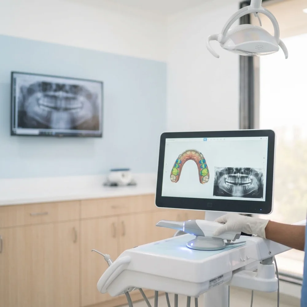
CBCT stands for cone beam computed tomography. In plain terms, it is a three-dimensional dental X-ray. Instead of a single flat image, it produces a full 3D picture of your teeth, jawbone, nerves and sinuses. Your dentist can rotate it on screen and look through it slice by slice, from any angle and in any plane. It gives depth and position, not just outline, and that is the whole reason it exists.
How does a CBCT scan work?
The scan itself is straightforward. You sit or stand still while a cone-shaped X-ray beam and a detector make a single rotation around your head, usually taking only a few seconds. During that one sweep the machine captures a series of images from many angles, and a computer combines them into a single three-dimensional volume. There is nothing to place inside your mouth, and you can breathe normally; the only thing asked of you is to stay still so the picture stays sharp.
What a CBCT shows that an ordinary X-ray cannot
A flat 2D X-ray, such as an RVG, flattens everything into one plane, which is fine for many questions but hides depth. A CBCT keeps the third dimension, so it can reveal things a normal X-ray simply cannot show clearly:
- The height, width and density of the bone, which matters a great deal before implant surgery
- The exact position of nerves, especially the nerve running through the lower jaw
- The position of the sinuses in the upper jaw, which sit close to the roots of the back teeth
- The true shape, number and curve of root canals, including hidden or unusual ones
- The precise position and angle of unerupted or impacted teeth, such as wisdom teeth
In each case the value is the same: the dentist is working from the actual anatomy rather than estimating it from a flat picture, which makes the treatment safer and more predictable.
When a CBCT scan is genuinely needed
A CBCT is a specialised tool for specific questions, not part of a routine visit. It earns its place when the extra detail will genuinely change what is done. Common, well-justified reasons include:
- Planning dental implants, where the height, width and density of the bone and the position of nearby nerves and sinuses all need to be known
- Assessing whether there is enough bone before a graft or a sinus procedure
- Difficult or impacted wisdom teeth, especially when a root sits close to the nerve in the lower jaw
- Some complex or repeat root canals, where the canals are curved, hidden or unusual in shape
- Investigating a suspected cyst, other jaw pathology, or an injury that a flat X-ray cannot fully show
When you do not need a CBCT
Just as usefully, plenty of dentistry does not call for one. A routine check-up, spotting decay between teeth, a simple filling, and most straightforward root canals are all handled well by an ordinary 2D digital X-ray. A CBCT gives more information than those situations require, at a higher radiation dose, so reaching for it by default would be the wrong instinct. If a simpler digital X-ray answers the question, that is what should be used. Whether your own case needs a 3D scan is a clinical judgement made after an examination, not a box ticked for every patient.
CBCT vs a regular dental X-ray, side by side
| At a glance | Regular dental X-ray (RVG) | CBCT scan |
|---|---|---|
| Dimension | Flat, two-dimensional image | Three-dimensional volume you can view from any angle |
| What it shows | Decay, root tips and bone level in a single plane | Bone height, width and density, nerve and sinus position, root canal shape and impacted teeth |
| Radiation dose | Very low, a small intraoral dose | Higher than a single small dental X-ray, but still low |
| When it is used | Routine check-ups, fillings, most root canals, spotting hidden decay | Planning implants, bone grafting, difficult wisdom teeth, complex root canals, suspected jaw pathology |
What about the radiation?
This is the part most people want answered plainly. A CBCT scan does use more radiation than a single small dental X-ray, because it captures a whole three-dimensional volume rather than one flat image. Even so, the dose stays low, and a dental CBCT is a focused scan of the jaws, not a whole-body medical CT. It is more than an everyday X-ray, and less than many people fear.
The sensible way to weigh it is the principle every good clinic follows: keep any dose as low as reasonably achievable, and take a CBCT only when the added detail will genuinely change the diagnosis or the plan. If a simpler 2D X-ray can answer the question, that is what should be used. Because seeing a nerve, a sinus or the exact bone before surgery can matter so much, a CBCT is often well justified in the specific situations above; for a routine check-up it is not, and it should not be taken out of curiosity. As with any X-ray, tell your dentist if you are, or might be, pregnant, and a scan that is not urgent can usually wait.
What to expect, and how we use it
Having a CBCT scan taken is quick and comfortable. You hold still for the few seconds it takes the scanner to rotate, and the 3D image is ready for your dentist to study shortly afterwards. At Prudent Dental Clinic in Viman Nagar, Pune, we use CBCT imaging when planning dental implants, so the position of an implant can be worked out from the actual bone and the nearby nerves and sinuses rather than estimated from a flat picture. You can read more about the imaging we use, or book an appointment if you are considering treatment that may call for a clearer 3D view.
Sources & further reading
American Dental Association (MouthHealthy) · NIDCR, US National Institute of Dental & Craniofacial Research · NHS, Dental Health · Indian Dental Association
CBCT 3D dental scans: your questions answered
Planning an implant or need a clearer 3D picture? Talk to us in Viman Nagar. Call +91 70287 22200.
Call +91 70287 22200 · Open 7 days, 9 AM–9 PM (5 PM Sun)