

Key takeaways
- RVG (radiovisiography) is simply a digital dental X-ray, a small sensor replaces the old film and the image appears on screen in seconds.
- It uses substantially less radiation than the old film system, commonly cited as up to 80–90% lower.
- It lets your dentist see hidden decay, infection, bone levels and root canals, and show you the image the same visit.
- The dose is very small, but a good dentist still only takes an X-ray when it will genuinely guide your treatment.
If your dentist has said they need to take an "RVG", it can sound more alarming than it is. An RVG is nothing more than a modern, digital version of a dental X-ray, and it is one of the most routine and useful tools in the clinic. Here is what it means, why it is done, and why the radiation involved is far less than most people assume.
What does RVG mean?
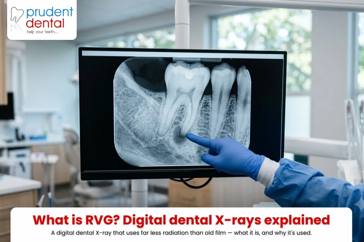
RVG stands for radiovisiography. In plain terms, it is a digital intraoral X-ray. A small electronic sensor, about the size of the old film packet, is placed inside your mouth next to the tooth being examined. A brief, low-dose X-ray is taken, and instead of a film that has to be developed in chemicals, the image appears almost instantly on the dentist's computer screen.
That immediacy is the whole point. Your dentist can turn the monitor towards you, point to the exact spot they are concerned about, and explain what they are seeing while you are still in the chair. There is no waiting, no darkroom, and the image can be brightened, enlarged and stored in your record.
Why do dentists take an RVG?
Because much of what matters in dentistry is hidden. The surfaces between your teeth, the inside of a tooth, the tip of a root and the bone that supports everything are all invisible to a visual examination. An RVG brings them into view. Common reasons include:
- Finding decay starting between teeth, before it becomes a large cavity
- Checking the extent of an infection or abscess at the root of a tooth
- Measuring the length and curve of root canals before a root canal treatment
- Assessing the bone level around teeth in gum disease
- Locating an impacted or wisdom tooth before removal
- Planning restorations, and checking existing fillings and crowns
In each case, the X-ray replaces guesswork with evidence, so the treatment plan is based on what is actually happening inside the tooth rather than an educated guess.
How is RVG different from an ordinary X-ray?
They rely on the same principle: a small X-ray beam passes through the tooth and is captured by a detector on the other side. The difference is the detector. A conventional dental X-ray uses a film that must be chemically processed, which takes longer and needs a higher dose to expose properly. RVG uses a reusable digital sensor.
That single change brings several benefits: the image is instant, no developing chemicals are involved, the radiation dose is lower, and the picture can be digitally enhanced and shared. It is worth noting that an RVG is a small, detailed image of one or a few teeth. It is different from a panoramic X-ray (OPG), which is taken from outside the mouth and shows the whole jaw in one wide view. Each has its place, and your dentist chooses based on what they need to see.
| RVG at a glance | Digital RVG X-ray | Traditional film X-ray |
|---|---|---|
| Detector | Small reusable electronic sensor | Film packet developed in chemicals |
| Image speed | Appears on screen within seconds | Takes longer, needs chemical processing |
| Radiation dose | Lower, commonly cited as up to 80 to 90 percent less | Higher dose to expose the film properly |
| Viewing the image | Can be enlarged, brightened and stored digitally | Fixed film image, no enhancement |
| Area shown | Detailed view of one or a few teeth | Same small intraoral area as RVG |
Is the radiation from an RVG safe?
This is the question most people really want answered, and it is a fair one. The honest answer is that the dose is very small. Digital dental sensors like RVG use substantially less radiation than the older film they replaced, commonly cited as up to 80 to 90 percent lower. The exposure from a single intraoral image is in the same broad range as the natural background radiation everyone is exposed to over a short stretch of ordinary life, from the sun, soil and the air around us.
That does not mean X-rays should be taken casually. Good practice follows a simple principle: keep every dose as low as reasonably achievable, and take an X-ray only when it will actually change the diagnosis or the treatment. A protective apron or thyroid collar may be used, and your dentist should be able to explain why a particular image is needed. If you are pregnant or think you might be, tell your dentist, who will usually postpone anything non-urgent.
Used this way, digital dental X-rays are considered a safe and essential part of diagnosis by dental and health authorities worldwide. The far greater risk is usually leaving a hidden problem, such as decay between teeth or an infection at a root tip, undiagnosed until it becomes painful and expensive.
What to expect at the clinic
Having an RVG taken is quick and painless. You will be asked to hold still while the sensor is positioned and a brief exposure is taken, and the image is on screen moments later. At Prudent Dental Clinic in Viman Nagar, Pune, digital imaging is part of a normal dental examination when it is needed, and you can see and discuss the findings in the same visit. You can read more about the digital X-rays we use, or book an appointment if something is troubling you.
Sources & further reading
Indian Dental Association · American Dental Association (MouthHealthy) · World Health Organization, Oral Health
RVG digital X-rays: your questions answered
Need a check-up in Viman Nagar? Book with Prudent Dental Clinic. Call +91 70287 22200.
Call +91 70287 22200 · Open 7 days, 9 AM–9 PM (5 PM Sun)